AI in Emergency Medicine
AI Fracture Detection in the ED: What the Evidence Supports
Why this matters
AI fracture tools make clinicians more sensitive in reader studies, and NICE allows four of them under conditions. No randomized trial has yet published patient outcomes. How to deploy one as a second reader.
Recommended next step
Pair this article with the free guide or course store if you want a more structured framework you can apply at the bedside or in leadership conversations.
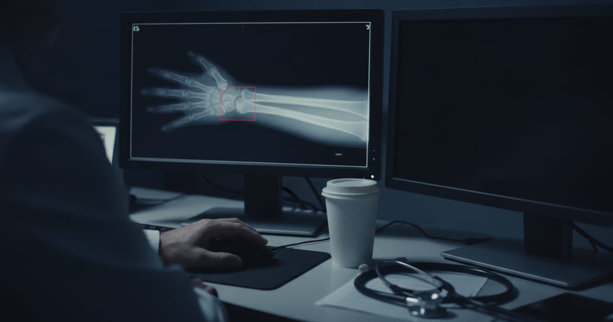
A 67-year-old woman who lives alone fell on her outstretched hand on the porch steps. Her wrist films look clean to you on both views. Then the AI fracture tool your department switched on last month posts a secondary image to PACS with a box drawn over the distal radius. You look again and still see nothing, and the final radiology read will not land until morning. She wants to go home tonight.
AI fracture detection makes clinicians more sensitive in reader studies, and that is the extent of the proof. No trial has yet shown that it changes what happens to patients, so the tool works best as a second reader with a local audit behind it.
What NICE recommended, and what it did not
In January 2025 the National Institute for Health and Care Excellence (NICE) published an early value assessment of AI fracture detection on X-rays in urgent care [1]. It assessed five products and allowed four. Rayvolve and TechCare Alert can be used for people of any age. BoneView and RBfracture can be used for people aged 2 years and over. A fifth, qMSK, was limited to research [1]. The guidance was later moved to a new reference number, HTG739, with the recommendations unchanged [2].
The permission is narrow. The tools can be used during a 2-year evidence generation period, after which the companies are expected to submit that evidence to NICE [1]. They can only be used while that evidence is being generated, and only once they have regulatory approval that includes NHS England's Digital Technology Assessment Criteria [1]. They are used in addition to standard care. NICE states that they do not replace the definitive radiology review and that "clear local protocols will need to be in place" [1]. NICE also notes that the tools may not suit some groups, including children and young people [1].
NICE's assessment puts missed or delayed fracture diagnosis on radiographs at around 3% to 10% of cases [1].
NICE advises the NHS in England. A US department buys under device clearance and its own review, on the same reader studies.
What the reader studies measured
A widely cited 2022 meta-analysis pooled 42 studies of AI reading on its own [3]. Pooled sensitivity on external validation sets was 91% for AI and 94% for clinicians, and pooled specificity was 91% and 94%. The differences were not statistically significant. Of the 42 studies, 22 (52%) were judged at high risk of bias [3]. The authors' conclusion was that AI and clinicians had comparable reported performance.
The question that matters more to an emergency physician is whether a clinician reads better with the tool. A 2026 meta-analysis of 26 studies answered it in aggregate: pooled clinician sensitivity rose from 77% to 87% with AI assistance, and specificity from 88% to 92%. Eight of the 26 studies were at high risk of bias [9]. Two widely cited multi-reader studies show how such numbers are produced.
- Guermazi 2022. Twenty-four readers, including emergency physicians, orthopedists, radiologists and physician assistants, read 480 examinations with and without AI [4]. Fracture prevalence was set at 50%. Per-patient sensitivity rose from 64.8% to 75.2%, specificity rose from 90.6% to 95.6%, and reading time fell by 6.3 seconds per examination. The sensitivity gain was significant in every body region except shoulder and clavicle (+4.2%) and spine (+2.6%) [4].
- Duron 2021. Six emergency physicians and six radiologists read radiographs from 600 adults at 17 French centres, with obvious fractures excluded [5]. AI raised sensitivity by 8.7 percentage points and specificity by 4.1, and cut false-positive fractures per patient by 41.9% in patients without fractures. The 15% reduction in reading time was not statistically significant [5].
Both are retrospective reader studies with case sets enriched for fractures, read by clinicians who knew they were being tested. Both list co-authors from Gleamer, the company that makes the software tested. An enriched case set raises the positive predictive value a reader sees. In a real ED queue, where most films are normal, a larger share of flagged boxes will be false.
The gaps: outcomes, children, and the wrong box
No randomized trial has yet published patient outcome results. A multicenter randomized trial in emergency departments in two countries finished recruiting in May 2025. Its preliminary conference abstract reported no numbers, and it warned that AI assistance may increase additional imaging and prolong ED treatment because of uncertainty about AI findings [10]. SAMURAI-fracture is a cluster randomized crossover trial in emergency departments and minor injury units across 4 NHS trusts, testing RBfracture in patients older than 2 years [6]. Sites switch the tool on and off month by month for 6 months. The primary outcome is "inappropriate healthcare contacts": unnecessary fracture clinic referrals, repeat attendances and recalls for missed fractures. Recruitment began in January 2026 and was planned to run 6 months at each site, so results are pending [6].
One real-world signal comes from a before-and-after study. A Milan emergency department added BoneView for orthopaedists working night shifts without an on-site radiologist [11]. Across 28,655 patients, fracture-related recalls were 26 before and 25 after, and missed fractures 16 before and 13 after. The change was not statistically significant [11].
Children are a separate question. A 2026 meta-analysis of 16 pediatric studies, 10,203 children and 21,789 radiographs found pooled sensitivity of 93% and specificity of 91%, and 93% and 88% on external validation [7]. The authors still called for paired-design studies with external geographic validation before the models are generalized [7]. Check the age limit of the specific product: two of the four NICE products are restricted to age 2 and over [1].
The third gap is the wrong box. None of the fracture studies above tested what a confidently wrong AI mark does to a reader. The closest evidence comes from mammography. In an experiment with 27 radiologists, inexperienced readers rated 79.7% of mammograms correctly when the purported AI suggestion was right and 19.8% when it was wrong. Very experienced readers fell from 82.3% to 45.5% [8]. Mammography is a different task, and the size of the effect will not transfer directly. The direction of the effect should be expected in fracture reads too.
Deploying it as a second reader
Four rules come out of that evidence.
- Read first, then open the overlay. In both reader studies, the AI marks were shown with the films, and neither study tested a read-first sequence. A first independent read is the one protection against the anchoring seen in the mammography experiment, and it costs seconds.
- Decide who owns a disagreement. When the box and the clinician disagree, the protocol needs a default: repeat views, splint and follow up, or ask for a radiology read before discharge. For the woman on the porch, a splint and orthopedic follow-up is safer than a discharge that rests on either reader alone.
- Audit both directions. Count the boxes the clinicians overruled that radiology confirmed, and the fractures radiology found that the tool missed. Review them monthly. That local number is what tells you whether the tool helps in your population.
- Match the product to your patients. Confirm the cleared age range and body regions before go-live. In the 480-examination reader study, spine and shoulder were the regions where the tool added the least [4].
The same discipline applies to any AI read in the department, as covered in running a new AI tool in shadow mode first and what to demand from a vendor's evidence packet. The AI ECG as a second reader follows the same pattern.
Key Takeaways
- NICE's January 2025 assessment lets four AI fracture tools be used for 2 years while evidence is gathered, in addition to standard care and never in place of the radiology review.
- With AI, reader sensitivity rose from 64.8% to 75.2% in a 480-examination study enriched to 50% fracture prevalence. Shoulder, clavicle and spine showed no significant gain.
- No randomized trial has published patient outcomes yet. One real-world before-and-after study found a non-significant drop in missed fractures, 16 to 13 in 28,655 patients.
- Read the film before you open the overlay, name an owner for disagreements, and audit both directions every month: boxes you overruled and fractures the tool missed.
FAQ
Does AI fracture detection replace the radiologist's read?
No. NICE states that the tools do not replace the definitive radiology review and are used in addition to standard care [1]. The reader studies tested AI as an aid to a clinician who still made the call.
How accurate is AI at finding fractures on X-rays?
In a 2022 meta-analysis of 42 studies, AI on its own and clinicians performed about the same, with pooled sensitivity of 91% for AI and 94% for clinicians on external test sets [3]. More than half of those studies were at high risk of bias. Across 26 studies of clinicians reading with AI help, pooled sensitivity rose from 77% to 87% [9].
Can AI fracture detection be used on children?
Some products can. NICE allows two of its four named products at any age and two from age 2 [1]. A 2026 pediatric meta-analysis reported pooled sensitivity of 93% and specificity of 91%, and its authors asked for more external validation before wide use [7].
Dr. Chet's Take
I have worked more than 25 years as an emergency department attending, and I have served as a TelEmergency medical director, where the clinician at the bedside and the reader of the study are often not in the same building. This article reaches the right conclusion. The reader studies are real, the effect on sensitivity is real, and randomized outcome evidence does not exist yet. Those three facts can sit together. The mistake is to let a sales deck turn the first two into a promise about the third. I have no quarrel with the technology. My quarrel is with the speed at which a conditional permission gets described as an endorsement once it reaches a purchasing meeting. A box on a radiograph is a reason to look again. It is not a diagnosis, and nobody should chart it as one.
That being said, I think the article understates where the tool may matter most. In a large academic department, a radiologist reads the film within hours. In a small rural ED, the preliminary read may be the only read for a long night, and the clinician reading it may not read many wrists in a month. The honest answer is that this setting has barely been studied, and that is the setting where a second reader could carry the most weight. It is also where an unaudited tool could do the most quiet harm, because nobody downstream catches the false reassurance until the patient comes back. The places most likely to benefit are the places least likely to have the staff to measure it. Share the audit across a health system so the small sites are not left to run it alone.
If you are leading an emergency department and a vendor is in your inbox, ask for the reader-study numbers by body region and by age. Then ask what the tool did to the false-positive rate at a site with your fracture prevalence, and get the answer in writing. Put a monthly discrepancy review on the calendar before go-live, not after the first complaint, and give one named physician the job of reading it. If you are the attending on shift, read the film first, every time, and document your own read before you open the overlay. Teach residents the same order from their first week. The overlay is a second opinion, and a second opinion only has value when there was a first one.
— Chester Shermer, MD, FACEP | Emergency Medicine, 25+ Years Clinical Experience | State Surgeon
Continue Your Training
If you're an emergency physician (or any clinician treating patients daily) trying to understand how AI will actually impact your clinical practice — not just the hype — I put together a free practical guide. You can download the AI in EM Survival Guide here.
Browse All Courses at Global MedOps Command
Relevant Reading on Global MedOps Command:
How to Avoid Becoming an AI Casualty — Dr. Shermer's guide to navigating AI tools in clinical and operational settings without compromising judgment or patient outcomes.
Emergency Department Efficiency Playbook — Practical systems for throughput, triage optimization, and operational efficiency.
Read more from Dr. Shermer on Medium
Connect with Dr. Shermer: LinkedIn — Chester "Chet" Shermer, MD, FACEP.
References
- National Institute for Health and Care Excellence. Artificial intelligence (AI) technologies to help detect fractures on X-rays in urgent care: early value assessment. Health technology evaluation HTE20. Published 14 January 2025. nice.org.uk
- National Institute for Health and Care Excellence. HealthTech guidance HTG739 (migrated from HTE20, recommendations unchanged). nice.org.uk
- Kuo RYL, Harrison C, Curran TA, et al. Artificial Intelligence in Fracture Detection: A Systematic Review and Meta-Analysis. Radiology. 2022;304(1):50-62. PubMed 35348381
- Guermazi A, Tannoury C, Kompel AJ, et al. Improving Radiographic Fracture Recognition Performance and Efficiency Using Artificial Intelligence. Radiology. 2022;302(3):627-636. PubMed 34931859
- Duron L, Ducarouge A, Gillibert A, et al. Assessment of an AI Aid in Detection of Adult Appendicular Skeletal Fractures by Emergency Physicians and Radiologists: A Multicenter Cross-sectional Diagnostic Study. Radiology. 2021;300(1):120-129. PubMed 33944629
- Novak A, Vaz J, Espinosa Morgado AT, et al. Systematic assessment of the medical utility of radiology and diagnostic Artificial Intelligence in fracture detection (SAMURAI-fracture): a protocol for a multicentre cluster-randomised controlled trial. BMJ Open. 2026;16(8):e120631. PubMed 42629153
- Ximenes GF, Costa ÁL, Leite LL, et al. Are Artificial Intelligence Models Reliable for Clinical Application in Pediatric Fracture Detection on Radiographs? A Systematic Review and Meta-analysis. Clin Orthop Relat Res. 2026;484(2):371-385. PubMed 40839831
- Dratsch T, Chen X, Rezazade Mehrizi M, et al. Automation Bias in Mammography: The Impact of Artificial Intelligence BI-RADS Suggestions on Reader Performance. Radiology. 2023;307(4):e222176. PubMed 37129490
- Qin H, Ding Y, Ju J, et al. Enhanced fracture detection on radiographs with AI assistance for clinicians: a systematic review and meta-analysis. Ann Med. 2026;58(1):2610079. PubMed 41467767
- Breitwieser M. AI in emergency fracture diagnosis: Unintended consequences and hidden costs – insights from a randomized controlled trial. German Congress of Orthopaedics and Traumatology (DKOU 2025), Berlin, October 2025. Conference abstract. publisso.de
- Albano D, Vignati G, D'Andrea S, et al. Fracture Detection on Bone Radiographs: The Impact of an AI Tool on Orthopaedic Night Shifts. J Imaging. 2026;12(6):252. PubMed 42346915
Related simulation training: EM-Sim · Books by Dr. Shermer
Keep reading
Related reading and your next step.
Ready to go further? Move from this article into structured training, scenario-based rehearsal, and more physician-written guidance.
Course
Translate the article into a repeatable framework
Use the physician-led course when you want a structured framework for evaluating AI tools, protecting clinical judgment, and leading implementation decisions.
Simulation
Practice the decision path under pressure
Use EM-Sim when you want scenario-based repetition that turns article-level insight into physician-facing emergency-medicine reps.
Blog
Browse more articles
Explore the full blog for more on AI in emergency medicine, then head to the course and simulation pages when you want the structured next step.
Related Articles
ED Management
Left Without Being Seen: What Emergency Physicians Can Actually Fix
Patients who leave without being seen come back more often, and the front end that drives most walkouts is the part an emergency physician can redesign. Here is what the evidence supports.
AI in Emergency Medicine
AI for ED Staffing: Forecasts Are Not Shift Plans
AI can forecast emergency department arrivals, but a patient count is not a staffing plan. Learn how to validate forecasts, keep human control, and test a safer workflow.
AI in Emergency Medicine
Before AI Changes ED Care, Run It in Shadow Mode
Before an AI model changes an emergency department workflow, run it in shadow mode. Here is what to measure, who owns the stop rule, and what should earn go-live.